This section is about the methods of overcoming the fact that in Radial Aplasia, the soft tissues on the radial side of the forearm are very short compared to the ulna bone, causing the typical bent “club” hand. To surgically correct the hand position, this needs to be addressed. There are different approaches to this, some (most?) of which I will discuss in this post.
I am going out on a limb here (no pun intended), given my educational/professional background. Yet I am sure that discussing this topic will interest some parents – it certainly interested us. We especially found the notion that the ulna would shortened questionable before learning more about the reasoning behind the approach.
While I don’t claim the following to be scientific, I tried my best to follow the science and deduct carefully. If you find anything incorrect or have suggestions, please let me know!
The current methods of overcoming differences in length
The limiting factor in correcting radial club hand are the soft tissues – the nerves and vessels – that have a fixed length, limiting the angle by which the hand can be straightened. The first method that comes to mind (and is still prevalent today) to overcome this problem is to “stretch” the soft tissues before the surgical intervention. This can be done by either utilizing an external fixator (“external fixation”) or by other means of distraction, e.g., splints/orthoses or casts. For Radializations, distraction via an external fixator worn for several months before surgery is a common approach. In some cases, external fixation is utilized after surgery to hold everything in place (Dr. Standards approach).
The other possibility is to shorten the bone to match the length of the soft tissues (Dr. Paley’s approach in the latest generation – Gen. 3 as of this writing – of Ulnarization techniques [2]).
Dr. Paley discusses lengthening vs. shortening in his Paper “Shortening: The orthopedic theory of relativity.” [1]
Distraction vs. Shortening - A Comparison
Distraction of soft tissues in general
Applies force that results in extra load on the cartilage of the joints (a connective tissue providing a smooth, lubricated surface for articulation with minimal friction) and growth plates [1].
Many splints only have a negligible effect (more about this below)
Requires a lot of time
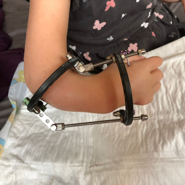
Distraction through external fixation
Tethers muscles and fascia [1].
Causes considerable strain on patients and parents.
Comes with a high risk of infections (primarily pin-site infections) that carry the risk of inhibiting the bone growth that is desperately needed in most cases of Radial Aplasia.
Extra surgeries to fix problems like ripped-out pins are not unusual.
Also, strong distraction over a relatively short period carries a risk of neurovascular stretch injury.
More information about external fixation can be found here.
Shortening of the ulna
Counterintuitively, shortening the ulna does not reduce the arm’s overall axial length (as it is limited by the length of the (short) soft tissues at the time of surgery) – this seems to be a common misconception. Also see [1].
Using locking plates and nails/K-wires (internal fixation) reduces the problems related to external fixation and makes rehabilitation easier and faster [1].
An osteotomy is – somewhat – a trauma like a fracture, and, in children, often results in bone overgrowth (well documented, for example, in fractures of the femoral shaft (thigh bone)). In many cases, this is a problem; in this particular case, it is an advantage, potentially recuperating some of the ulna length lost.
Right hand in self-made orthosis at around 9 months, after 6 months of wearing self-made orthoses at night.
For a perfect Ulnarization resulting in the maximum axial arm length combined with all the other benefits of Paley’s Ulnarization procedure (see “Surgical Methods” for more details), soft tissues would be distracted to (almost) match the length of the ulna before surgery, thus reducing the amount of shortening needed to a minimum,
without utilizing an external fixator, thus avoiding the drawbacks discussed above
avoiding the issues associated with distraction in general by distracting slowly over a long period to prevent neurovascular injury, ergo applying only little force, reducing the extra load on the cartilage of the joints and growth plates.
So, what about splinting? As already mentioned above, many regular splints have little to no effect as they primarily fix an already attainable hand position. If done poorly, one could splint forever, and the wrist would only be assured of staying the same. This is, for example, precisely what we want to achieve once the surgical correction has been done: locking the forearm in its position and avoiding any movement. It is clear by now that regular splinting, as Dr. Paley told us, “neither helps nor harms.”
On the other hand, the Ponseti method – see the digression – proves that successful deformity corrections can be achieved via external force introduction over an extended period. The difference is that a Ponseti brace is usually rigid (and sometimes not removable, e.g., a cast), holding the affected limb firmly in a position it was “forced” into. This level of fixation is unpracticable on the upper limbs and hardly achievable with the typical thermoplastic splints shown in the pictures. From my perspective, it is generally much more challenging to fixate small, fragile wrists than fixate two feet on a “snowboard-with-bindings”-like brace.
Digression: The Ponseti Method
Some research on the topic inevitably leads to the Ponseti method – a non-surgical treatment approach developed by Dr. Ignacio Ponseti in the 1940s for treating clubfoot. Based on gradual manipulation of the foot position in the affected baby or toddler, it involves several phases that are typically carried out over several weeks or months, starting with gentle pressure and traction techniques, later progressing to the application of plaster casts and special braces. The initial step involves gently bringing the clubfoot into an improved position, usually performed manually (and painlessly for the child).
Once the foot position has been corrected as much as possible, a plaster cast is applied to hold the foot in the new position. The cast is regularly changed, typically every one to two weeks, to continue correcting the clubfoot. After several weeks or months of treatment with plaster casts, the child is provided with a special brace known as a Ponseti brace. This brace is worn at night and supports the corrected foot position to prevent the recurrence of the clubfoot.
The Ponseti method has proven to be highly successful in correcting clubfeet in babies and toddlers and is still applied worldwide. In most cases, it allows for complete restoration of foot function and average walking ability without the need for surgery.
Typical splint for the night (this one was made by professionals in Hamburg) of low-temperature thermoplastic material, heated to around 60-70°C in water and then shaped around the extended arm, maintaining the shape upon cooling
Orthosis of soft silicone made by a professinal orthoses maker. Did not fit very well and left hands sweaty. Impedes the sensoric stimulation crucial for a child's development.
Shaping Orthoses
The question for me was: how do I make a splint (I use the words splint and orthesis synonymously) that applies force when worn at night but can be removed during the day? It needed to be soft, not to introduce too much force, and be in the desired shape of the forearm/wrist, not the current one.
I then thought of Invisalign, a company that uses a digital approach for correcting dental misalignments. They use digital models of the teeth in their current and desired positions, and then 3D-print dental aligners can gradually move the teeth toward the desired positions. Patients replace the aligners with the next set every one to two weeks. The more extensive the correction, the more steps are required. Without digital models and 3D printing, this procedure would be nearly impossible.
The idea now was – not new in orthopedics – to capture the shape of the forearms, wrists, and hands using a 3D scanner, then make slight corrections on the computer and create a positive mold for the orthoses through 3D printing. The mold would be used to craft the orthoses via vacuum molding. The resulting orthoses, made from a soft thermoplastic material, should exert a constant gentle pressure in the desired direction while being worn. Once pressure is no longer applied (as the soft tissues have lengthened), a new iteration for further correction is designed and manufactured.
The images below show the Ulnarization procedure by Dr. Paley with osteotomy on the left and three “Stages” of soft tissue distraction as I envisioned it when designing orthoses on the computer. Unfortunately, we never got x-rays done with orthoses shortly before surgery.
A 3D-Scan of the (stretched) right hand (at around 9 Months)
Steps of the Ulnarization procedure. Source: Paley D. Shortening: The orthopedic theory of relativity. J Limb Lengthen Reconstr 2020;6:1-4.
Stage 1. Own depiction, based on: Paley D. Shortening: The orthopedic theory of relativity. J Limb Lengthen Reconstr 2020;6:1-4.
Stage 2. Own depiction, based on: Paley D. Shortening: The orthopedic theory of relativity. J Limb Lengthen Reconstr 2020;6:1-4.
Stage 3. Own depiction, based on: Paley D. Shortening: The orthopedic theory of relativity. J Limb Lengthen Reconstr 2020;6:1-4.
As Dr. Paley’s Paper is published under the CC BY-NC-SA license, all depictions derived from it are also under said license.
Building Orthoses & Conclusion
I consequently developed, built, and experimented with (here, my educational background came in handy) a wide variety of orthoses of different materials and stiffness, some including spring mechanisms, etc. (see pictures). You can find a detailed description of the process of making these orthoses and a discussion of the findings in this section.
After consulting our daughter’s orthopedist and physiotherapist, as well as an orthoses manufacturer, she would wear the best versions every night. In 2021, we showed the orthoses to Dr. Paley in Warsaw.
Conclusion:
In our case, we believe that consistently wearing a customized orthosis every night and soft bandages during the day resulted in
Our daughter benefiting from relatively straight yet mobile hands during the time before surgery, making everyday activities easier and less change to adapt to after Ulnarization.
The osteotomy performed during Ulnarization being in the millimeters, not centimeters.
Ultimately, this resulted in her forearms being a bit longer (how much is impossible to say, but I would guess about one to 1,5 centimeters, considering that 2-3cm ulna shortening are typical), thus requiring less lengthening.
Was it worth the considerable effort? Absolutely.
While one instance does not allow for general conclusions (our daughter’s soft tissues might have lengthened due to other, unknown factors), comparison of her hands (range of motion – “ROM” – immediately before surgery) with untreated TAR-syndrome club hands (thumbs present) showed a striking difference.
If you decide to go down the same path as we did, start by talking to your child’s orthopedist and find an experienced orthosis builder. If you choose to take things into your own hands, consult a qualified professional before attempting any measures!
Making of a CAD-modeled custom orthosis. Many iterations are necessary to find the perfect target shape
Experimental orthosis with feeler gauge leave as spring mechanism.
Experimental orthosis with feeler gauge leave as spring mechanism.
PLA padded with feeler gauge leave
Natural hand position
PLA unpadded with feeler gauge leave
Newborn right hand with radial aplasia
Right baby hand radial aplasia (few months of age) – palmar view
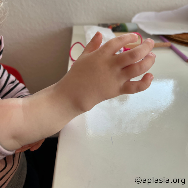
Bandaged hand is very straight – a few months beore Ulnarization
Right hand one year after Ulnarization
3D positive models for the vacuum molding of orthoses, early stage and late stage.
Below are 3D CAD renderings of an early-stage and a late-stage positive model for vacuum molding.
Deszczyński, Jarosław & Albrewczyński, Tomasz & Shannon, Claire & Paley, Dror. (2021). Radial Club Hand Treated by Paley Ulnarization Generation 3: Is This the New Centralization?. Children. 8. 562. 10.3390/children8070562.
(1) Background: Patients treated with the two previous generations of ulnarization developed a bump related to the ulnar head becoming prominent on the radial side of the hand. To finally remedy this problem, a third generation of ulnarization was developed to keep the ulnar head contained. While still ulnar to the wrist center, the center of the wrist remains ulnar to the ulnar head, with the ulnar head articulating directly with the trapezoid and when present the trapezium. (2) Methods: Between 2019 and 2021, 22 radial club hands in 17 patients were surgically corrected with this modified version of ulnarization. (3) Results: In all 17 patients, the mean HFA (hand–forearm-angle) correction was 68.5° (range 12.2°–88.7°). The mean ulna growth was 1.3 cm per year (range 0.2–2 cm). There were no recurrent radial deviation deformities more than 15° of the HFA. (4) Conclusions: This new version of ulnarization may solve the problem of the ulna growing past the carpus creating a prominent ulnar bump. The results presented are preliminary but promising. Longer-term follow-up is needed to fully evaluate this procedure.
Paley, Dror. (2018). Limb reconstruction in the early 21 st century: The indications are broader and wider. Journal of Limb Lengthening & Reconstruction. 4. 65. 10.4103/2455-3719.253397.
Paley, Dror. (2017). The Paley ulnarization of the carpus with ulnar shortening osteotomy for treatment of radial club hand. SICOT-J. 3. 5. 10.1051/sicotj/2016040.
Recurrent deformity from centralization and radialization led to the development in 1999 of a new technique by the author called ulnarization. This method is performed through a volar approach in a vascular and physeal sparing fashion. It biomechanically balances the muscle forces on the wrist by dorsally transferring the flexor carpi ulnaris (FCU) from a deforming to a corrective force. The previous problems of a prominent bump from the ulnar head and ulnar deviation instability were solved by acutely shortening the diaphysis and by temporarily fixing the station of the carpus to the ulnar head at the level of the scaphoid. This is the first report of this modified Paley ulnarization method, which the author considers a significant improvement over his original procedure..