Surgical methods
On this page, you will find a condensed overview of the standard surgical procedures to treat Radial Aplasia. The information is based on our own research, scientific papers, answers from surgeons of four different hand-surgical centers of expertise (we asked many questions, and so should you), other parents of affected children (we are deeply grateful to all of them), and online sources. Please also read our Disclaimer!
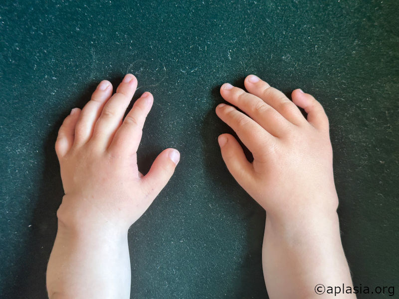
Ulnarization
There is a detailed (including medical illustration) explanation of the procedure online.
The main differences to the Radialization procedure are:
- No distraction of tissue is necessary before moving the bone
- Incisions are done on the palmar side so the surgeons can see the nerves during the procedure
- The FCU (flexor carpus ulnaris) is transferred to help as a corrective force (the muscle that is on the ulnar side and is transferred to the radial side).
- The ulna bone is shortened (typically betweeen 2 – 3cm) to fit the “pocket” under the hand to hold position and continue growing in the right direction, and straightened at the same time (by removing a trapezoidal bone segment).
- No long-term splinting after treatment (only temporary night splints for a couple of weeks)
The main advantages of Ulnarization:
- No recurrence
- More strength
- Very mobile “wrist”
- Ability to bear weight on wrists
- No external fixator (Paley) with a higher risk of infections
- Two surgeries in 3 months (instead of 5 surgeries ranging over 1,5 years), fewer anesthesias (with resulting risks)
- Both hands are operated on simultaneously
- There is no need for year-long splinting until skeletal maturity has been reached
Significant differences between Ulnarizations (Dr.Paley / Dr. Standard):
According to our information – Dr. Standard does not regularly shorten the ulna and uses an external fixator to keep bone position fixated after Ulnarization (info from 2021) – this means that there has to be an external fixation device on the arm after the Ulnarization procedure (in contrast to Radialization, where the external fixator is used to do the soft-tissue distraction before re-positioning the bone).
Side Note: Thumbs
In TAR-Kids, the thumb is usually present, so we cannot comment on any experiences with pollicization. However, as TAR-Kids often have weaker thumbs, Dr. Paley and Dr. Shannon might perform a transfer of the FCR tendon to the thumb extensor. This transfer allows for more extension of the thumb.


Radialization
The Radialization procedure was invented by Dieter Buck-Gramko, and this knowledge was passed on to the surgeons currently working in Hamburg. The Radialization procedure is offered in many parts of Europe, although Centralizations are still performed in some places.
The Radialization procedure usually consists of two steps:
- A distraction phase (fixator for 2-4 months)
- The actual Radialization procedure: re-positioning of the bone (internal wires for 4-6 months)
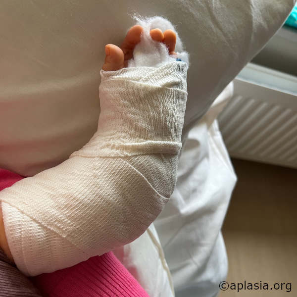
Distraction phase for Radialization
The distraction phase usually takes around three months, depending on the degree of radial deviation and soft tissue. It starts with surgery to
fit an external fixator device. After that, the child has one arm in the external fixator, and the parents must regularly adjust some screws to distract the soft tissue. This adjustment happens four times daily (for a certain amount of millimeters to be distracted). When the hand is in a straight position after several weeks, the external fixator stays on for another couple of weeks to hold this straight position. After that, the hand is ready for the Radialization procedure.
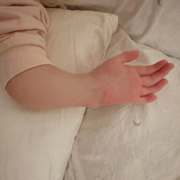
Radialization procedure
After the distraction phase, the hand is ready for the subsequent surgery, in which the ulna is moved into a more central position (“central position” does not imply equivalence with the Centralization procedure!).
After removing the external fixator, the ulna bone is repositioned towards the center of the hand / second metacarpal bone. The ulna bone is then fixated with K-wires, and the arm is splinted in a firm splint to protect the internal wires (so they don’t break in case the kids fall on them). The entire process entails two to three surgeries to correct one arm. The internal wires are removed after 3-6 months. When the surgeons remove wires on one side, they usually start with the other hand and put the fixator on in the same surgery. The entire process needs at least five surgeries (for two hands) and takes more than a year of continuous procedures for the child.

Here is an overview of what the surgical scheme looks like for the entire Radialization process:
- First surgery: right hand – putting on the external fixator.
- Second surgery: right hand – removing the external fixator, performing the Radialization procedure, and fixating with internal wires, the arm is put in a cast-like splint for several months.
- Third surgery: right hand – removal of internal wires; left hand – putting on the external fixator.
- Fourth surgery: left hand – performing the Radialization procedure and fixating with internal wires, the arm is put in a cast-like splint for several months.
- Fifth surgery: left hand – removal of internal wires.
Centralization
Some clinics still offer Centralization – the predecessor of the Radialization procedure. Centralization has many disadvantages, mainly because recurrence is high. If you have Centralization surgery as your only option, contact us and connect with other parents who have had a Centralization experience.
Relevant Sources and Publications
A comparison of different surgical procedures on Dr. Paley's website.
An overview of the different procedures on the website of Dr. Standard with an explanation of ulnarization by Dr. Standard (an older version of ulnarization where an external fixator is used to hold the position after moving the bone to the right position).
Paley, Dror. (2020). Shortening: The orthopedic theory of relativity. Journal of Limb Lengthening and Reconstruction. 6. 1. 10.4103/2455-3719.288573.
Paley, Dror. (2018). Limb reconstruction in the early 21 st century: The indications are broader and wider. Journal of Limb Lengthening & Reconstruction. 4. 65. 10.4103/2455-3719.253397.
Paley, D., Robbins, C.A. (2015). Case 91: Ulnarization as Treatment for Radial Clubhand (RCH). In: Rozbruch, S., Hamdy, R. (eds) Limb Lengthening and Reconstruction Surgery Case Atlas. Springer, Cham. https://doi.org/10.1007/978-3-319-18020-5_250
Buck-Gramcko D. Radialization as a new treatment for radial club hand. J Hand Surg Am. 1985 Nov;10(6 Pt 2):964-8. doi: 10.1016/s0363-5023(85)80013-7. PMID: 4078287.
Paley, Dror. (2017). The Paley ulnarization of the carpus with ulnar shortening osteotomy for treatment of radial club hand. SICOT-J. 3. 5. 10.1051/sicotj/2016040.
Recurrent deformity from centralization and radialization led to the development in 1999 of a new technique by the author called ulnarization. This method is performed through a volar approach in a vascular and physeal sparing fashion. It biomechanically balances the muscle forces on the wrist by dorsally transferring the flexor carpi ulnaris (FCU) from a deforming to a corrective force. The previous problems of a prominent bump from the ulnar head and ulnar deviation instability were solved by acutely shortening the diaphysis and by temporarily fixing the station of the carpus to the ulnar head at the level of the scaphoid. This is the first report of this modified Paley ulnarization method, which the author considers a significant improvement over his original procedure.
Deszczyński, Jarosław & Albrewczyński, Tomasz & Shannon, Claire & Paley, Dror. (2021). Radial Club Hand Treated by Paley Ulnarization Generation 3: Is This the New Centralization?. Children. 8. 562. 10.3390/children8070562.
(1) Background: Patients treated with the two previous generations of ulnarization developed a bump related to the ulnar head becoming prominent on the radial side of the hand. To finally remedy this problem, a third generation of ulnarization was developed to keep the ulnar head contained. While still ulnar to the wrist center, the center of the wrist remains ulnar to the ulnar head, with the ulnar head articulating directly with the trapezoid and when present the trapezium. (2) Methods: Between 2019 and 2021, 22 radial club hands in 17 patients were surgically corrected with this modified version of ulnarization. (3) Results: In all 17 patients, the mean HFA (hand–forearm-angle) correction was 68.5° (range 12.2°–88.7°). The mean ulna growth was 1.3 cm per year (range 0.2–2 cm). There were no recurrent radial deviation deformities more than 15° of the HFA. (4) Conclusions: This new version of ulnarization may solve the problem of the ulna growing past the carpus creating a prominent ulnar bump. The results presented are preliminary but promising. Longer-term follow-up is needed to fully evaluate this procedure.